silvia
Joined: 10 Oct 2021
Posts: 259
Location: UK


|
 Posted: Oct 31, 2022 15:53 Post subject: My aim is to produce a photo that will have high-resolution characteristics Posted: Oct 31, 2022 15:53 Post subject: My aim is to produce a photo that will have high-resolution characteristics |
|
|
| At https://www.mineral-forum.com/message-board/viewtopic.php?p=79712#79712 Carles Millan wrote: | | silvia wrote: | We have many specimens that just will not behave when they see the camera. They look great to the eye but they look too average when a photo is taken. It is not an easy task as what works well with one specimen does not always work well with another.
|
Silvia, believe me, you are not alone. I know what I'm talking about. |
| At https://www.mineral-forum.com/message-board/viewtopic.php?p=79034#79034 silvia wrote: |
...new Canon camera (EOS R6 MARK II) using an EF-EOS M Mount adapter and a Canon EFM 28mm f/3.5Macro IS STM lens on an Joby tripod. We used the photo bracketing function to take multiple shots (35) that were processed using Canon’s Digital Photo Professional V4.16.10.0 software. All photos were depth composited before editing.
|
Thank you Carles you have opened up the introduction to a new and related topic for me.
My partner says I am too much of a perfectionist. I try to get every feature of the specimen in focus using the “aperture priority” technique, but it is not easy with minerals that have mirror like features. My aim is to produce a photo that will have high-resolution characteristics even when ‘blown up’ to poster size.
General Procedure
Camera: CANON EOS M6 MARK II
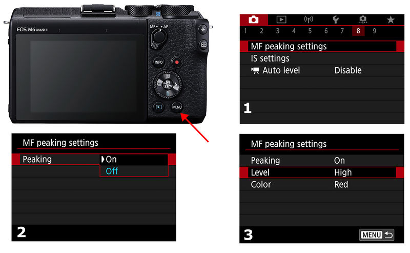
Use Aperture priority mode with camera set in manual. Activate the manual peaking mode. In this mode there will be a red line (or blue or yellow each set by the user) or a series of dots indicating which parts of the specimen are in focus. Note that not all parts of the specimen will be in focus, just those that show the red dots or the red line. As the focusing ring of the camera is slowly turned different parts of the specimen will display a series of red dots indicating that this part (or region) of the specimen is in focus. The dots or line will move from bottom to top (or vice-versa) of the specimen. I start at the bottom and lowly rotate the focusing ring – take a shot – then rotate the ring a little more then take another shot. I use the magnifying glass function of the camera as it enables me to magnify the portion of the specimen, albeit a small portion, I wish to become the main subject of the focus of a given shot. When complete, I have several dozen shots to process. The shots are processed with the Canon Photo Professional Version - 4 software, using the depth compositing tool.
This procedure works in about 98% of all photos taken, especially with specimens in the cabinet to Museum size. I find small specimens, miniature size or less, pose no real problems even when they have mirror-like features.
| Mineral: | - |
| Description: |
|
| Viewed: |
19050 Time(s) |

|
|
|


